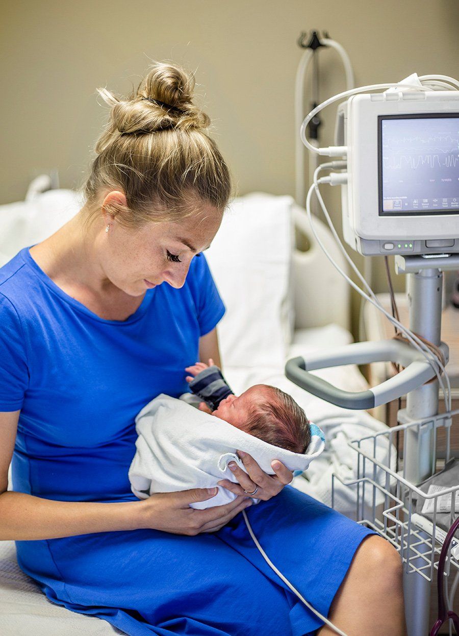
The Core Matrix: Why are babies born tight?
The Core Matrix: why are babies born tight?
The Womb Experience
As the embryo grows, and the fascial network evolves, so too does the complexity of support and connectivity of that fascial matrix. At first internal growth and differentiation may take precedence. Later, the size limitations, shape or position of the womb may be most significant for the developing fetus and the blueprint of the fetus’s connective tissue architecture, the fascial matrix.
Fascia (a form of connective tissue) surrounds every muscle, bone, organ, nerve, and vessel down to the cellular level. This connective tissue forms a three-dimensional web or matrix that connects from head to toe, arm to arm, without interruption.
This system’s function is to support vital organs in their correct positions and provide cohesions to all the body structures. The fascial matrix keeps everything separate yet interconnected at the same time.
Everything in the body is supported in connective tissue. Within that tissue is a network of nerves, blood vessels, organs, bones, and muscles. When an area of connective tissue is compressed, restricted, or compromised, the structure that is wrapped within the restricted fascia is also impeded.
The way the embryo/fetus lies in the uterus determines the ultimate pattern of the spine as well as the blueprint of the entire connective tissue arrangement. Whether the head or shoulders is to the right or left, or how the arms and legs are curled or twisted during development, are crucial factors in the final architecture of the infant.
As it grows, the embryo/fetus expands in size but retains the early pattern of position. Around the sixth month of pregnancy, the size limitation of the uterus or position of maternal pelvis creates new demands and challenges in the internal environment for the growing fetus.
The more severe this limitation within the uterus, the greater the likelihood for the fetus to rotate, fold, twist, and compress further into adaptations. By the time the fetus is ready to be born, he/she has developed a blueprint of connective tissue pulls that reflect intrauterine stresses and constraints.
We have found these intrauterine stresses consist of and are not limited to placenta previa, uterine bands, multiple pregnancies, bicornuate uterus, nuchal cord, oligohydramnios, and maternal scarring from prior surgeries.
The Journey from Inside to Outside
Although birth is a natural and wondrous event, the fetus faces further compressions, pulls, and twists to the connective tissue structure during the birth event.
Historically, the stresses of the birth experience are accepted as “normal” or “that is just the way it is”; however, the process in which a fetus can navigate his/her birth creates a lifelong effect on the infant’s structure.
There are many situations that occur during labor and delivery that further affect the fetus’s structure.
Under normal conditions, the woman’s pelvis reshapes itself to accommodate birth. If this reshaping process is interrupted by anesthesia, an epidural injection, the reshaping of the pelvis that normally occurs is inhibited.
When labor does not progress because the pelvic involvement is suppressed, Pitocin is introduced to force the uterus to contract artificially. The effect that this has on the fetus is like using the cranium as a battering ram to force the maternal pelvis to reshape to accommodate it.
Furthermore, when the fetal cranium passes through the birth canal, it’s very malleable and takes the form of the maternal pelvic structure. If you ram the fetus’s head into the maternal pelvis, these distortions end up becoming imprinted and locked into the infant’s cranium, as well as into the rest of the connective tissue structure.
Other significant stressors on the infant’s structure include and are not limited to vacuum-assisted birth, forceps-assisted birth, shoulder dystocia, nuchal cord, cesarean section, abnormally fast birth, and long difficult births. These birth conditions all distort and imprint in the weave of the connective tissue fibers.
Birth is an extraordinary event. The infant is experiencing a total change of environment and has no established ways of dealing with this change. New stimuli, such as what occurs during labor and delivery, can cause traumatic sensory overload that can induce structural contractions and distortions that may lock into the connective tissue of the newborn baby.
Connective tissue distortions, pulls, and restrictions are subtle, but significant disturbances to the function of the structures and organs. If left untreated, these distortions and pulls imprint and become imbedded into the connective tissue causing a child to compensate around the problem.
Since connective tissue is found everywhere in the body, these restrictions can interfere with the function of any nerve, muscle, organ, bone, or vessel within the body.
Immediately after birth, an infant may be unable to feed, may have digestive issues such as projectile vomiting or spitting up, or may have breathing issues. As these structural compensations remain in the body, they may interfere with developmental milestones such as creeping and crawling, standing, walking, and speech. The child must literally work with the restrictions that have already been established within the body from their embryonic blueprint and intrauterine stresses.
The direct effects of the compression of the connective tissue and structure of the fetal/newborn body from the in utero or birth experiences are a matter of concern. Contrary to common view, the infant will not grow out of these compressions and restrictions, but instead will grow into structural and functional limitations. These limitations become imprinted and locked into the fascial structure causing compensations and distortions over time.
FMCM is a gentle, non-surgical method that evaluates and corrects connective tissue restrictions, caused by in utero and/or labor and delivery compressions that occur to an infant’s structure.
We are incredibly passionate about working with families after the birth of their child and encourage treatments to begin as soon as is appropriate for the situation. We find that as time passes, the compressions that occur from in utero/labor and delivery, become more solidified in the tissue. While we can still eliminate the restrictions with FMCM techniques later in childhood, it is always easier to work with the imbalanced tissue as soon as possible after the trauma or stressful experience.
FMCM promises to improve the standard for pediatric care. This method has been proven to advance the functional health for newborns born prematurely and full term.
Michael and Kristen Myers, LMT, Copyright November 25, 2018
ABOUT
Fascial Matrix Connection Method® and The Matrix in Motion Somatic Movement Therapy is intended to serve as an adjunct to medically supervised healthcare. This article is not designed for and does not provide medical advice. All content in this article is for general information purposes only. The content of this article is not intended to be a substitute for professional medical or mental advice or care. You should consult with a healthcare provider for diagnosis and collaborative treatment. Michael Myers, Kristen Myers, and the Myers Institute ® are not responsible for any adverse effects or consequences resulting from the method discussed within the information of this article.
__________________________
Rosen, M. The trauma of Birth 2016


